Bionanointerfaces

Cells of all types take cues from the surfaces they encounter in their environment. The cells extract any or all of the mechanical, chemical, spatial, and even temporal information from the surface, but which features they read and how they integrate and translate them into specific behavioral responses is still almost a black box. 

Projects

Vascularized self-lubricating surfaces

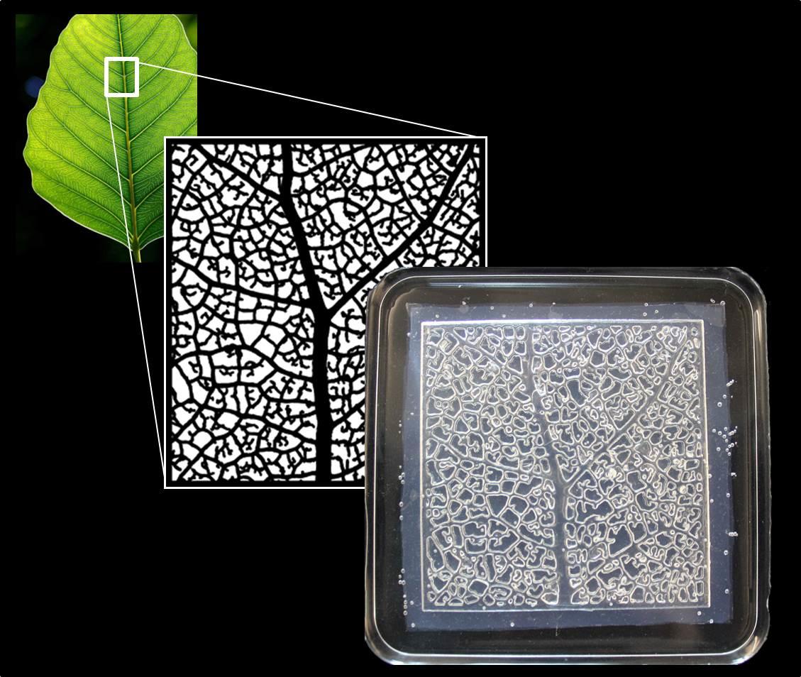
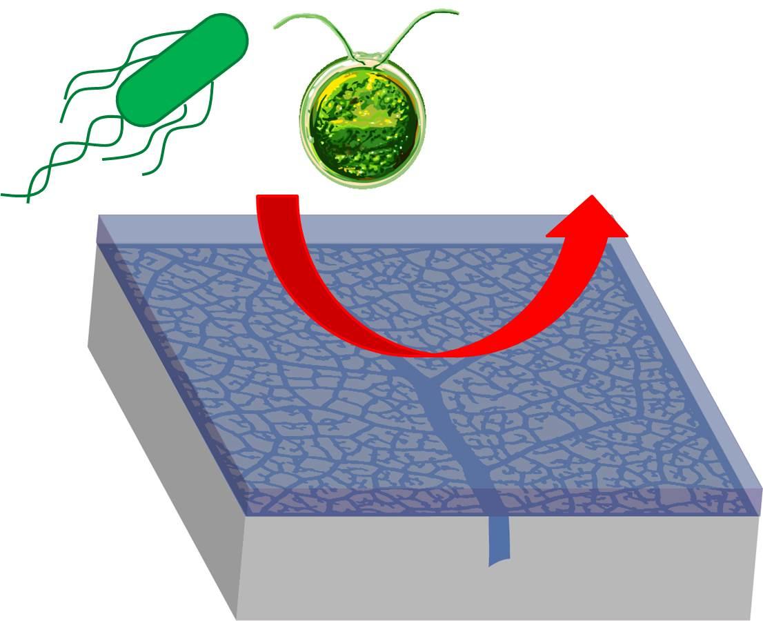
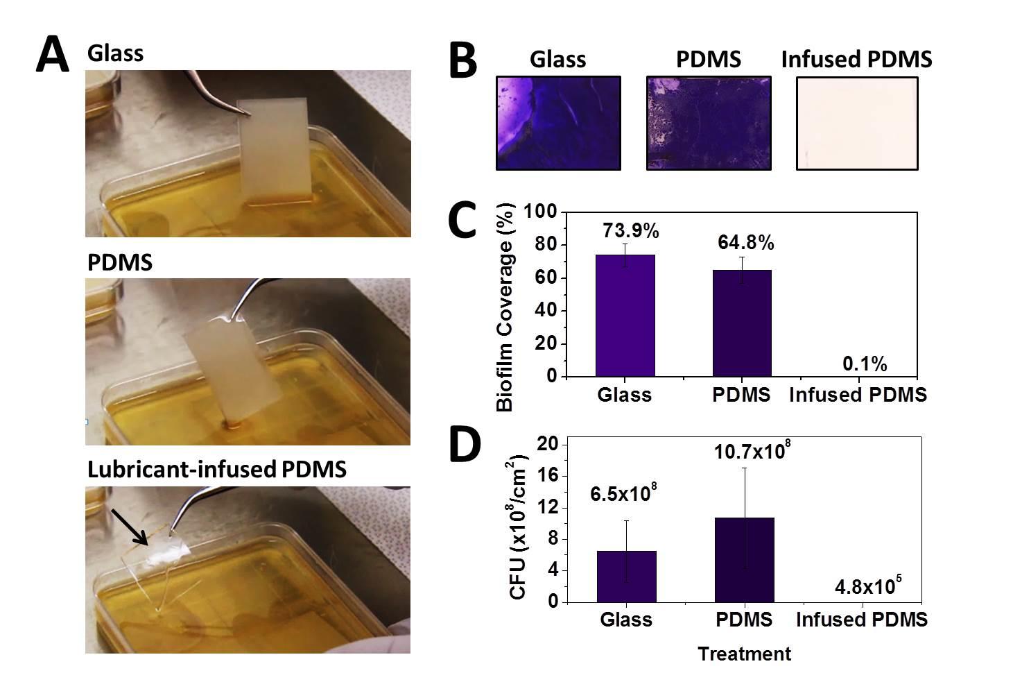
Fig. 1: Bio-inspired vascularized fouling-release surfaces.
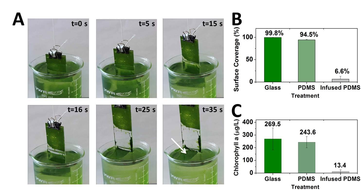
Fig. 2: Bio-inspired vacularized fouling-release surfaces.
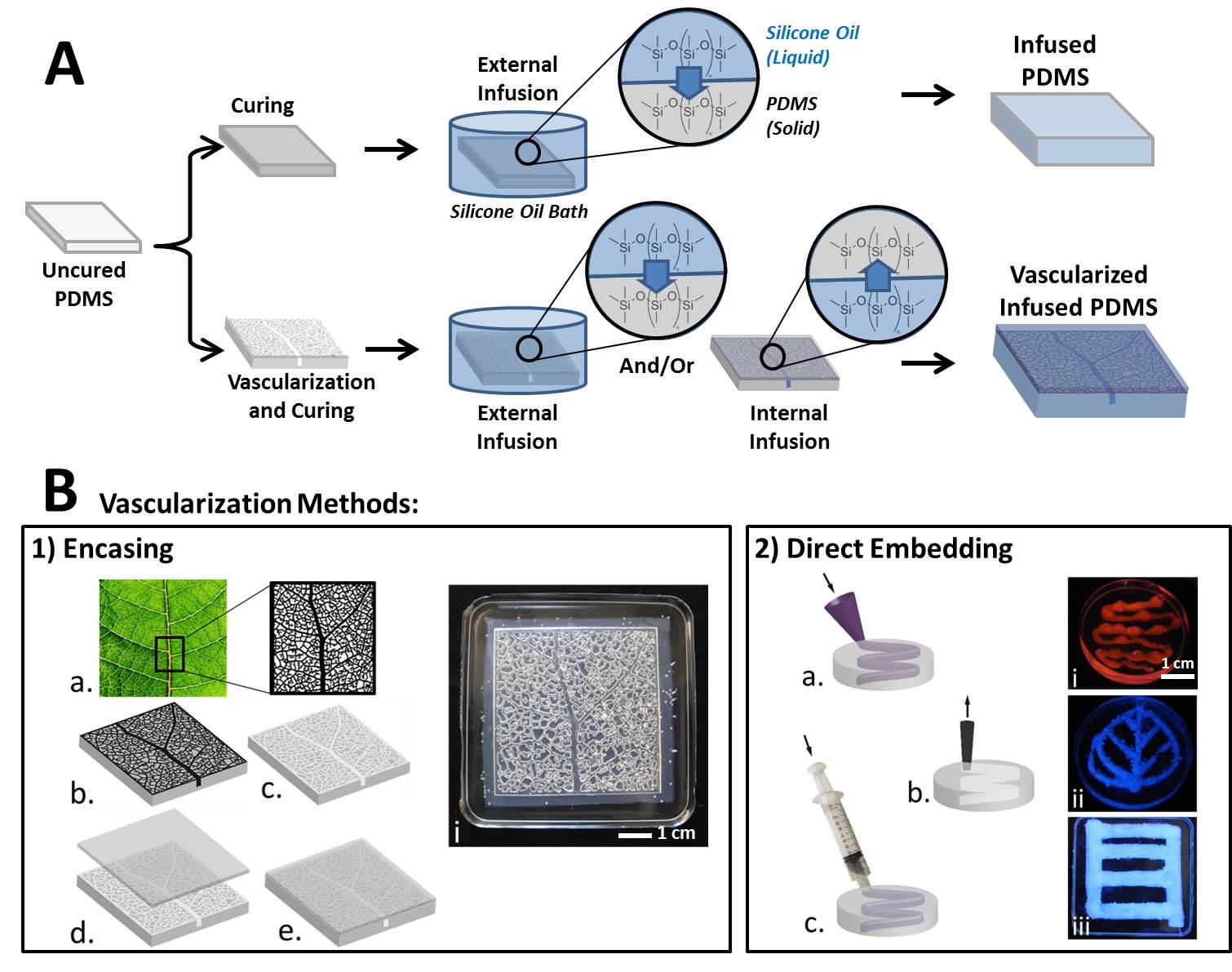
Fig. 3: (A) Schematic of the process to make either infused PDMS or vascularized, infused PDMS. For simple infused PDMS (upper row), cured PDMS is placed in a bath of silicone oil which diffuses into the PDMS solid. For vascularized PDMS, the vascular pattern is created before curing. The sample is then infused externally with silicone oil in the same manner as the non-vascularized PDMS, or internally through filling the vascular network, or both. (B) Methods of creating vascular networks within PDMS: 1) An encased network is created using a 3D mold (a) to create the pattern in PDMS (b). The mold is removed from the cured PMDS (c) and the pattern is covered with a second sheet of PDMS (d and e) which is chemically bonded to the pattern using plasma. (i) an image of a 3D leaf vasculature network after encasing. 2) An embedded network is created following the procedures developed by Lewis et al. (citation). (a) A pattern of 20% w/w pluronic F127 gel at 25 °C is embedded in uncured PDMS. (b) The PDMS is allowed to cure, cooled to 4 °C, and the liquid pluronic is evacuated. (c) The channel is refilled with silicone oil. (i) An image of fluorescently dyed PDMS in a hand-drawn sinuous channel, (ii) a hand-drawn leaf-shape network, and (iii) a 3D-printed linear network.
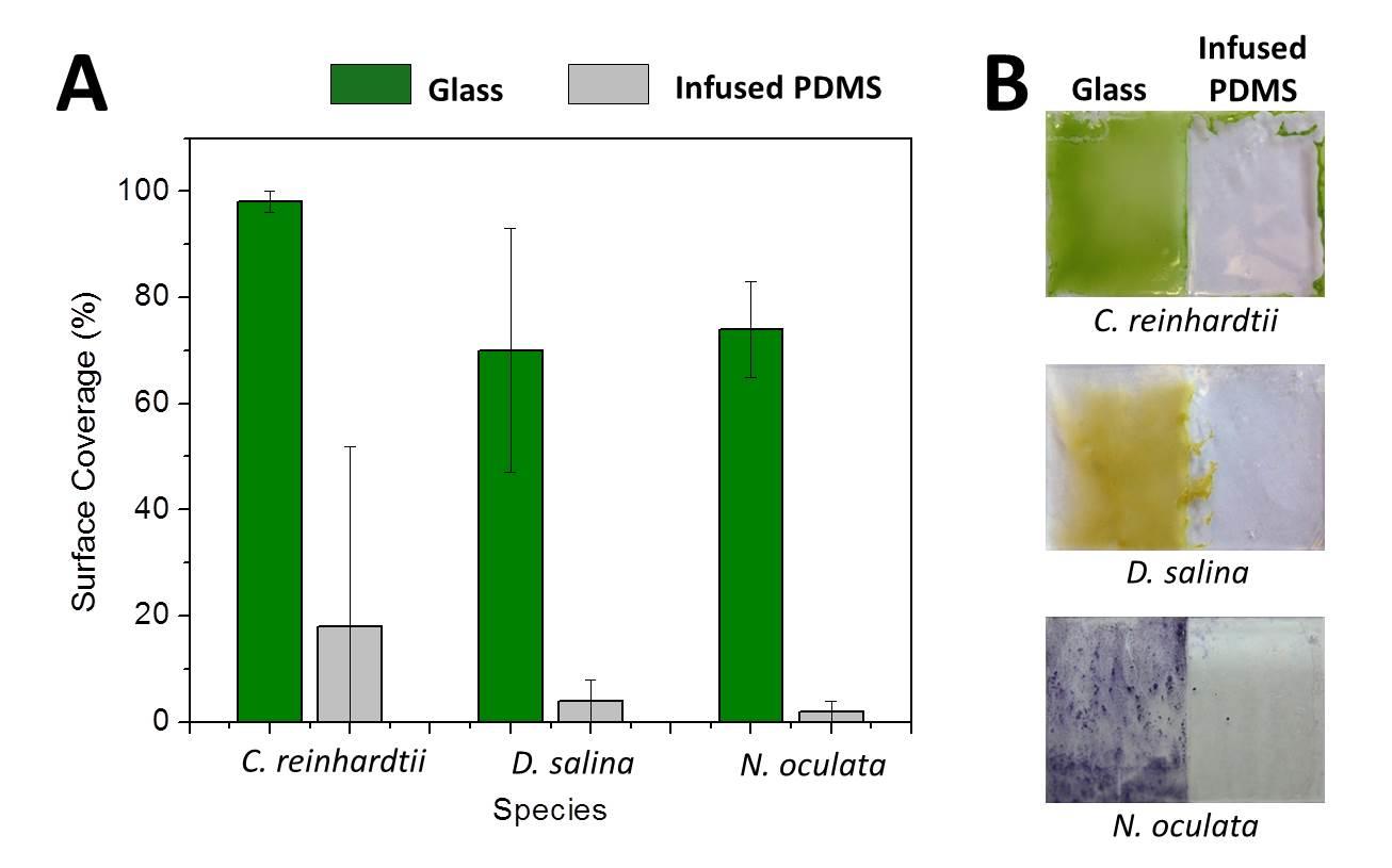
Fig. 6: (A) Surface coverage of biofilm remaining on lubricant-infused PDMS versus glass control surfaces after incubation in and removal from the liquid medium containing C. reinhardtii, D. salina, or N. oculata. For all three species, there was a significant reduction in the amount of biofilm remaining on the SLIPS surfaces (P < 0.05). (B) Images of the substrates used in the analysis.

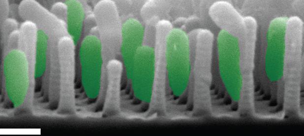
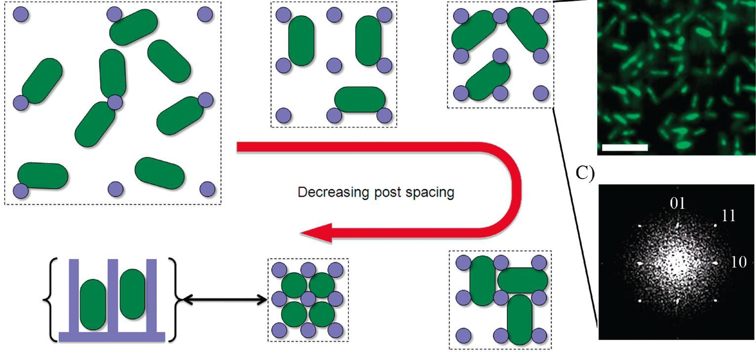
We have recently developed a technique for fabricating nanostructured surfaces that enables us to choose and independently vary any combination of geometry, spacing, stiffness, and chemistry, and are using it to analyze how surface features direct bacterial assembly, mammalian cell development, and marine invertebrate settlement at a level of systematic detail that was previously not possible.

As our insight into the cell-nanosurface interface develops, the same fabrication technique will enable us to design and construct surfaces that encode instructions for cell behavior for purposes ranging from anti-biofilm coatings to neural chips to organ development scaffolds.

Amini S, Kolle S, Petrone L, Ahanotu O, Sunny S, Sutanto CN, Hoon S, Cohen L, Weaver JC, Aizenberg J, et al. Preventing mussel adhesion using lubricant-infused materials. Science. 2017;357 (6352) :668-673. Publisher's VersionAbstract

Mussels are opportunistic macrofouling organisms that can attach to most immersed solid surfaces, leading to serious economic and ecological consequences for the maritime and aquaculture industries. We demonstrate that lubricant-infused coatings exhibit very low preferential mussel attachment and ultralow adhesive strengths under both controlled laboratory conditions and in marine field studies. Detailed investigations across multiple length scales—from the molecular-scale characterization of deposited adhesive proteins to nanoscale contact mechanics to macroscale live observations—suggest that lubricant infusion considerably reduces fouling by deceiving the mechanosensing ability of mussels, deterring secretion of adhesive threads, and decreasing the molecular work of adhesion. Our study demonstrates that lubricant infusion represents an effective strategy to mitigate marine biofouling and provides insights into the physical mechanisms underlying adhesion prevention.

Alvarez MM, Aizenberg J, Analoui M, Andrews AM, Bisker G, Boyden ES, Kamm RD, Karp JM, Mooney DJ, Oklu R, et al. Emerging Trends in Micro- and Nanoscale Technologies in Medicine: From Basic Discoveries to Translation. ACS Nano. 2017;11 (6) :5195-5214. Publisher's VersionAbstract

We discuss the state of the art and innovative micro- and nanoscale technologies that are finding niches and opening up new opportunities in medicine, particularly in diagnostic and therapeutic applications. We take the design of point-of-care applications and the capture of circulating tumor cells as illustrative examples of the integration of micro- and nanotechnologies into solutions of diagnostic challenges. We describe several novel nanotechnologies that enable imaging cellular structures and molecular events. In therapeutics, we describe the utilization of micro- and nanotechnologies in applications including drug delivery, tissue engineering, and pharmaceutical development/testing. In addition, we discuss relevant challenges that micro- and nanotechnologies face in achieving cost-effective and widespread clinical implementation as well as forecasted applications of micro- and nanotechnologies in medicine.

Sutton A, Shirman T, Timonen JVI, England GT, Kim P, Kolle M, Ferrante T, Zarzar LD, Strong E, Aizenberg J. Photothermally triggered actuation of hybrid materials as a new platform for in vitro cell manipulation. Nat. Commun. 2017;8 :14700. Full TextAbstract

Mechanical forces in the cell’s natural environment have a crucial impact on growth,
differentiation and behaviour. Few areas of biology can be understood without taking into account how both individual cells and cell networks sense and transduce physical stresses. However, the field is currently held back by the limitations of the available methods to apply physiologically relevant stress profiles on cells, particularly with sub-cellular resolution, in controlled in vitro experiments. Here we report a new type of active cell culture material that allows highly localized, directional and reversible deformation of the cell growth substrate, with control at scales ranging from the entire surface to the subcellular, and response times on the order of seconds. These capabilities are not matched by any other method, and this versatile material has the potential to bridge the performance gap between the existing single cell micro-manipulation and 2D cell sheet mechanical stimulation techniques.

Kovalenko Y, Sotiri I, Timonen JVI, Overton JC, Homes G, Aizenberg J, Howell C. Bacterial Interactions with Immobilized Liquid Layer. Adv. Healthcare Mater. 2017;6 (15) :1600948. Publisher's VersionAbstract

Bacterial interactions with surfaces are at the heart of many infection-related problems in healthcare. In this work, the interactions of clinically relevant bacteria with immobilized liquid (IL) layers on oil-infused polymers are investigated. Although oil-infused polymers reduce bacterial adhesion in all cases, complex interactions of the bacteria and liquid layer under orbital flow conditions are uncovered. The number of adherent Escherichia coli cells over multiple removal cycles increases in flow compared to static growth conditions, likely due to a disruption of the liquid layer continuity. Surprisingly, however, biofilm formation appears to remain low regardless of growth conditions. No incorporation of the bacteria into the layer is observed. Bacterial type is also found to affect the number of adherent cells, with more E. coli remaining attached under dynamic orbital flow than Staphylococcus aureus, Pseudomonas aeruginosa under identical conditions. Tests with mutant E. coli lacking flagella confirm that flagella play an important role in adhesion to these surfaces. The results presented here shed new light on the interaction of bacteria with IL layers, highlighting the fundamental differences between oil-infused and traditional solid interfaces, as well as providing important information for their eventual translation into materials that reduce bacterial adhesion in medical applications.

Hu Y, You J-O, Aizenberg J. Micropatterned Hydrogel Surface with High-Aspect-Ratio Features for Cell Guidance and Tissue Growth. ACS Appl. Mater. Interfaces. 2016;8 (34) :21939-21945.Abstract

Surface topography has been introduced as a new tool to coordinate cell selection, growth, morphology, and differentiation. The materials explored so far for making such
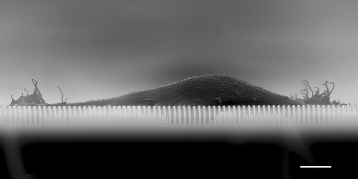
structural surfaces are mostly rigid and impermeable. Hydrogel, on the other hand, was proved a better synthetic media for cell culture because of its biocompatibility, softness, and high permeability. Herein, we fabricated a poly(2-hydroxyethyl methacrylate) (pHEMA) hydrogel substrate with high-aspectratio surface microfeatures. Such structural surface could effectively guide the orientation and shape of human mesenchymal stem cells (HMSCs). Notably, on the flat hydrogel surface, cells rounded up, whereas on the microplate patterned hydrogel surface, cells elongated and aligned along the direction parallel to the plates. The microplates were 2 μm thick, 20 μm tall, and 10−50 μm wide. The interplate spacing was 5−15 μm, and the intercolumn spacing was 5 μm. The elongation of cell body was more pronounced on the patterns with narrower interplate spacing and wider plates. The cells behaved like soft solid. The competition between surface energy and elastic energy defined the shape of the cells on the structured surfaces. The soft permeable hydrogel scaffold with surface structures was also demonstrated as being viable for longterm cell culture, and could be used to generate interconnected tissues with finely tuned cell morphology and alignment across a few centimeter sizes.

Juthani N, Howell C, Ledoux H, Sotiri I, Kelso S, Kovalenko Y, Tajik A, Vu TL, Lin JJ, Sutton A, et al. Infused polymers for cell sheet release. Scientific Reports. 2016;6 (1) :26109. Full TextAbstract

Tissue engineering using whole, intact cell sheets has shown promise in many cell-based therapies. However, current systems for the growth and release of these sheets can be expensive to purchase or difficult to fabricate, hindering their widespread use. Here, we describe a new approach to cell sheet release surfaces based on silicone oil-infused polydimethylsiloxane. By coating the surfaces with a layer of fibronectin (FN), we were able to grow mesenchymal stem cells to densities comparable to those of tissue culture polystyrene controls (TCPS). Simple introduction of oil underneath an edge of the sheet caused it to separate from the substrate. Characterization of sheets post-transfer showed that they retain their FN layer and morphology, remain highly viable, and are able to grow and proliferate normally after transfer. We expect that this method of cell sheet growth and detachment may be useful for low-cost, flexible, and customizable production of cellular layers for tissue engineering.

Friedlander RS, Vogel N, Aizenberg J. Role of Flagella in Adhesion of Escherichia coli to Abiotic Surfaces. Langmuir. 2015;31 (22) :6137-6144. Full TextAbstract

Understanding the interfacial activity of bacteria is of critical importance due to the huge economic and public health implications associated with surface fouling and biofilm formation. The complexity of the process and difficulties of predicting microbial adhesion to novel materials demand study of the properties of specific bacterial surface features and their potential contribution to surface attachment. Here, we examine flagella, cell appendages primarily studied for their cell motility function, to elucidate their potential role in the surface adhesion of Escherichia coli - a model organism and potential pathogen. We use self-assembled monolayers (SAMs) of thiol-bearing molecules on gold films to generate surfaces of varying hydrophobicity, and measure adhesion of purified flagella using quartz crystal microbalance. We show that flagella adhere more extensively and bind more tightly to hydrophobic SAMs than to hydrophilic ones, and we propose a two-step vs a single-step adhesion mechanism that accounts for the observed dissipation and frequency changes for the two types of surfaces, respectively. Subsequently, study of the adhesion of wild-type and flagella knockout cells confirms that flagella improve adhesion to hydrophobic substrates, whereas cells lacking flagella do not show preferred affinity to hydrophobic substrates. Together, these properties bring about an interesting ability of cells with flagella to stabilize emulsions of aqueous culture and dodecane, not observed for cells lacking flagella. This work contributes to our overall understanding of nonspecific bacterial adhesion and confirms that flagella, beyond motility, may play an important role in surface adhesion.

Chen J, Howell C, Haller CA, Patel MS, Ayala P, Moravec KA, Dai E, Liu L, Sotiri I, Aizenberg M, et al. An immobilized liquid interface prevents device associated bacterial infection in vivo. Biomaterials. 2017;113 :80-92.Abstract

Virtually all biomaterials are susceptible to biofilm formation and, as a consequence, device-associated infection. The concept of an immobilized liquid surface, termed slippery liquid-infused porous surfaces (SLIPS), represents a new framework for creating a stable, dynamic, omniphobic surface that displays ultralow adhesion and limits bacterial biofilm formation. A widely used biomaterial in clinical care, expanded polytetrafluoroethylene (ePTFE), infused with various perfluorocarbon liquids generated SLIPS surfaces that exhibited a 99% reduction in S. aureus adhesion with preservation of macrophage viability, phagocytosis, and bactericidal function. Notably, SLIPS modification of ePTFE prevents device infection after S. aureus challenge in vivo, while eliciting a significantly attenuated innate immune response. SLIPS-modified implants also decrease macrophage inflammatory cytokine expression in vitro, which likely contributed to the presence of a thinner fibrous capsule in the absence of bacterial challenge. SLIPS is an easily implementable technology that provides a promising approach to substantially reduce the risk of device infection and associated patient morbidity, as well as health care costs.

Sunny S, Cheng G, Daniel D, Lo P, Ochoa S, Howell C, Vogel N, Majid A, Aizenberg J. Transparent antifouling material for improved operative field visibility in endoscopy. Proc. Nat. Acad. Sci. 2016;113 (42) :11676-11681.Abstract

Inspection devices are frequently occluded by highly contaminating fluids that disrupt the visual field and their effective operation. These issues are particularly striking in endoscopes, where the diagnosis and treatment of diseases are compromised by the obscuring of the operative field by body fluids. Here we demonstrate that the application of a liquid-infused surface coating strongly repels sticky biological secretions and enables an uninterrupted field of view. Extensive bronchoscopy procedures performed in vivo on a porcine model shows significantly reduced fouling, resulting in either unnecessary or ∼10–15 times shorter and less intensive lens clearing procedures compared with an untreated endoscope.

Camera-guided instruments, such as endoscopes, have become an essential component of contemporary medicine. The 15–20 million endoscopies performed every year in the United States alone demonstrate the tremendous impact of this technology. However, doctors heavily rely on the visual feedback provided by the endoscope camera, which is routinely compromised when body fluids and fogging occlude the lens, requiring lengthy cleaning procedures that include irrigation, tissue rubbing, suction, and even temporary removal of the endoscope for external cleaning. Bronchoscopies are especially affected because they are performed on delicate tissue, in high-humidity environments with exposure to extremely adhesive biological fluids such as mucus and blood. Here, we present a repellent, liquid-infused coating on an endoscope lens capable of preventing vision loss after repeated submersions in blood and mucus. The material properties of the coating, including conformability, mechanical adhesion, transparency, oil type, and biocompatibility, were optimized in comprehensive in vitro and ex vivo studies. Extensive bronchoscopy procedures performed in vivo on porcine lungs showed significantly reduced fouling, resulting in either unnecessary or ∼10–15 times shorter and less intensive lens clearing procedures compared with an untreated endoscope. We believe that the material developed in this study opens up opportunities in the design of next-generation endoscopes that will improve visual field, display unprecedented antibacterial and antifouling properties, reduce the duration of the procedure, and enable visualization of currently unreachable parts of the body, thus offering enormous potential for disease diagnosis and treatment.

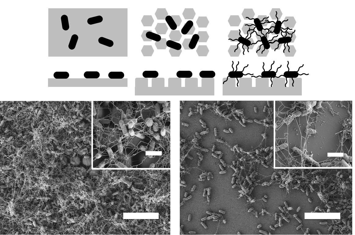
Friedlander RS, Vlamakis H, Kim P, Khan M, Kolter R, Aizenberg J. Bacterial flagella explore microscale hummocks and hollows to increase adhesion. Proc. Nat. Acad. Sci. 2013;110 (14) :5624-5629. Publisher's VersionAbstract
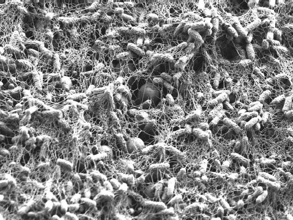
Biofilms, surface-bound communities of microbes, are economically and medically important due to their pathogenic and obstructive properties. Among the numerous strategies to prevent bacterial adhesion and subsequent biofilm formation, surface topography was recently proposed as a highly nonspecific method that does not rely on small-molecule antibacterial compounds, which promote resistance. Here, we provide a detailed investigation of how the introduction of submicrometer crevices to a surface affects attachment of Escherichia coli. These crevices reduce substrate surface area available to the cell body but increase overall surface area. We have found that, during the first 2 h, adhesion to topographic surfaces is significantly reduced compared with flat controls, but this behavior abruptly reverses to significantly increased adhesion at longer exposures. We show that this reversal coincides with bacterially induced wetting transitions and that flagellar filaments aid in adhesion to these wetted topographic surfaces. We demonstrate that flagella are able to reach into crevices, access additional surface area, and produce a dense, fibrous network. Mutants lacking flagella show comparatively reduced adhesion. By varying substrate crevice sizes, we determine the conditions under which having flagella is most advantageous for adhesion. These findings strongly indicate that, in addition to their role in swimming motility, flagella are involved in attachment and can furthermore act as structural elements, enabling bacteria to overcome unfavorable surface topographies. This work contributes insights for the future design of antifouling surfaces and for improved understanding of bacterial behavior in native, structured environments.
MacCallum N, Howell C, Kim P, Sun D, Friedlander R, Ranisau J, Ahanotu O, Lin JJ, Vena A, Hatton B, et al. Liquid-Infused Silicone As a Biofouling-Free Medical Material. ACS Biomater. Sci. Eng. 2015;1 (1) :43-51.Abstract
There is a dire need for infection prevention strategies that do not require the use of antibiotics, which exacerbate the rise of multi- and pan-drug resistant infectious organisms. An important target in this area is the bacterial attachment and subsequent biofilm formation on medical devices (e.g., catheters). Here we describe nonfouling, lubricant-infused slippery polymers as proof-of-concept medical materials that are based on oil-infused polydimethylsiloxane (iPDMS). Planar and tubular geometry silicone substrates can be infused with nontoxic silicone oil to create a stable, extremely slippery interface that exhibits exceptionally low bacterial adhesion and prevents biofilm formation. Analysis of a flow culture of Pseudomonas aeruginosa through untreated PDMS and iPDMS tubing shows at least an order of magnitude reduction of biofilm formation on iPDMS, and almost complete absence of biofilm on iPDMS after a gentle water rinse. The iPDMS materials can be applied as a coating on other polymers or prepared by simply immersing silicone tubing in silicone oil, and are compatible with traditional sterilization methods. As a demonstration, we show the preparation of silicone-coated polyurethane catheters and significant reduction of Escherichia coli and Staphylococcus epidermidis biofilm formation on the catheter surface. This work represents an important first step toward a simple and effective means of preventing bacterial adhesion on a wide range of materials used for medical devices.
  •  
  • 1 of 2
  • »

Clinging to crevices, E. coli thrive, Harvard press release, April 10, 2013.